- Operation Hours: Mon-Fri (8:30am-5:30pm), Sat (8:30am-12:30pm), Closed on Sun & Public Holiday
- +65 6235 7888
Positron Emission Tomography – Computer Tomography (PET-CT) scan.
What is a PET-CT scan?
PET-CT imaging is a combination of metabolic, molecular, anatomical
and structural imaging. Whilst conventional CT and MRI focuses more
on anatomical and structural information, PET-CT imaging provides
additional information regarding the metabolic and molecular
changes that occur as a result of a disease process.
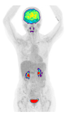
Prior to a PET-CT scan. Radioactive tracers are injected intravenously.
These radioactive tracers enable the attending Nuclear Medicine Physician
to evaluate specific metabolic or molecular changes that characterizes a
certain disease process.
Advanced Medicine imaging boasts two of the first
truly digital PET/CTs in Singapore. Being truly digital,
we are able to perform PET-CT scans at a lower radiation
dose and a shorter scanning time without compromising on
lesion detectability. In addition, this state-of-the-art
technology enables the detection of smaller lesions, a
difficult task for older analogue PET-CT scanners.
PET-CT can be performed for the following indications:
• Diagnosis and staging of cancer.
• Monitoring of treatment response.
• Detection of cancer recurrence.
• In fever of unknown origin.
• In tumor-induced osteomalacia.
• Cardiac imaging.
• Assessment of an underlying neurodegenerative disorder.
Currently the following PET radiotracers are offered at Advanced Medicine Imaging:
18F-Fluorodeoxyglucose (FDG).
FDG is the most commonly utilized radiotracer in PET-CT imaging.
Increase glucose utilization is seen in many disease processes,
for example, cancer, infection and inflammatory conditions.
Certain conditions can result in a decreased glucose metabolism,
for example, neurodegenerative disorders (eg, Alzheimer’s disease).

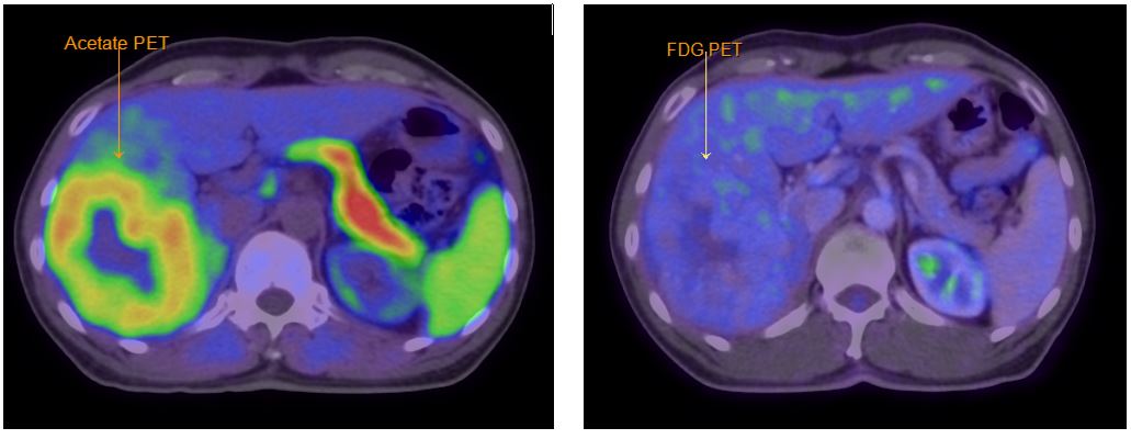
C11-Acetate
Acetate PET-CT has been utilized in different types of cancer eg, renal cell
neoplasm, myeloma, prostate cancer and hepatocellular carcinoma (HCC). In our centre,
Acetate PET-CT is used mainly in the evaluation of hepatocellular carcinoma (HCC). This
is sometimes performed in conjunction with a FDG PET-CT.
Acetate PET-CT usually detects the less aggressive well differentiated tumor whilst FDG PET-CT detects
the more aggressive less differentiated tumor components. This dual tracer approach helps identify
the different tumor clones as well as in improving lesion detection.
68 Gallium-PSMA PET-CT
PSMA is a transmembrane protein over expressed in approximately 90% of prostate cancers.
This radiotracer is commonly utilized and is the most accurate imaging agent currently available
for the staging, restaging and response assessment of prostate cancer. PSMA PET-CT is also used
in the work-up of patients prior to 177Lu-PSMA radionuclide therapy


68 Gallium-Dotatate PET-CT
Ga-68 Dotatate is a PET radiotracer that has high affinity for type 2 somatostatin receptors (SSTR).
Certain cancers, for example well differentiated neuro-endocrine tumors, paragangliomas, meningiomas
and mesenchymal tumors express type 2 SSTRs and can be imaged on DOTATATE PET-CT.


18F-Fluoroethyltyrosine (FET) PET-CT
18F-Fluoroethyltyrosine (FET) is an amino acid radiotracer which enter cells via the system L amino acid
transporter. It is mainly used in brain tumor imaging as an adjunct to MRI. It aids in identifying tumor
extent, in guiding biopsy as well as in treatment monitoring.

For more details, please contact Advanced Medicine Imaging at 6708-7888 or email imaging@dvancedmedicine.sg. We will be happy to assist you further.