
Imaging Centre
- IMAGING
- INTRODUCTION
- IMAGING SERVICES
- PATIENT PREPARATION
Introduction
01
The Imaging Centre at Asia HealthPartners is an integral part of our commitment to providing the best in healthcare. The latest imaging technology available at the Imaging Center ensures that the best clinical outcomes are achieved, from diagnosis to treatment.
To enhance service quality and provide seamless medical care, we also offer a filmless digital environment for image viewing, reporting, as well as storage with the Picture Archiving and Communication System (PACS). This allows viewing of images from a remote site as well as storage of patient studies for future follow-up comparisons.
Watch our video below to understand more on imaging scans.
Technology
Asia HealthPartners Milestones
- 2006 - 5th site globally to acquire the GE Signa® HDxt, the first high definition MRI scanner in Asia Pacific
- 2007 - 1st site in Singapore to acquire the GE LightSpeed™ VCT XT, the first low dose CT heart scan in the world
- The CT system is faster and gives higher resolution images compared to previous generation scanners.
- 2011 - First in Singapore to acquire VEO™ CT750 HD with the latest low dose technology
Services
02
Digital X-rays
Ultrasound Studies
Digital Mammography
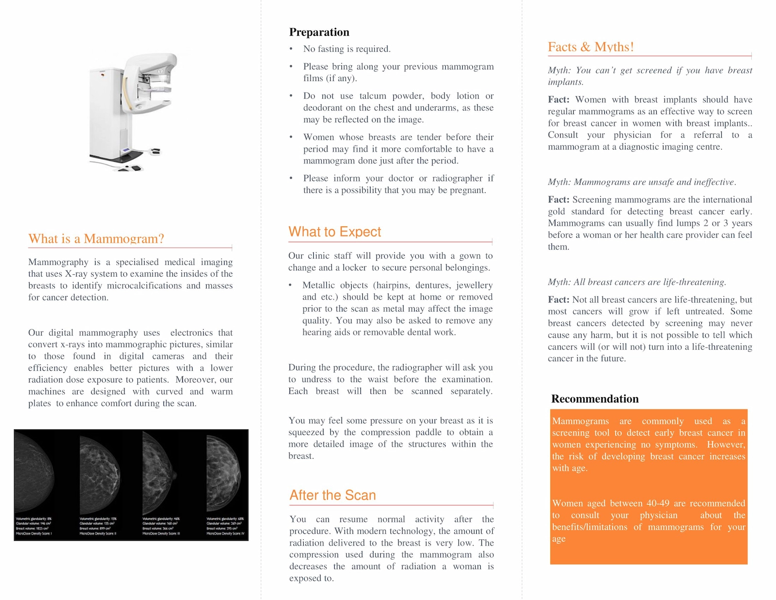
Digital mammography is a specialised medical imaging that uses x-ray system to examine the insides of the breasts to identify microcalcifications and masses for cancer detection. Our digital mammography uses electronics that convert x-rays into mammographic pictures, which are similarly found in digital cameras and their efficiency enables better pictures with a lower radiation dose exposure to patients.
Moreover, our machines are designed with curved and warm plates to enhance comfort during the scan. As the risk of developing breast cancer increases with age, women between 40-49 years of age are recommended to consult your physician about the benefits/limitations of mammograms for your lifestyle.
Computed Tomography (CT Scan) and CT Angiogram (CTA) [128-Slice]
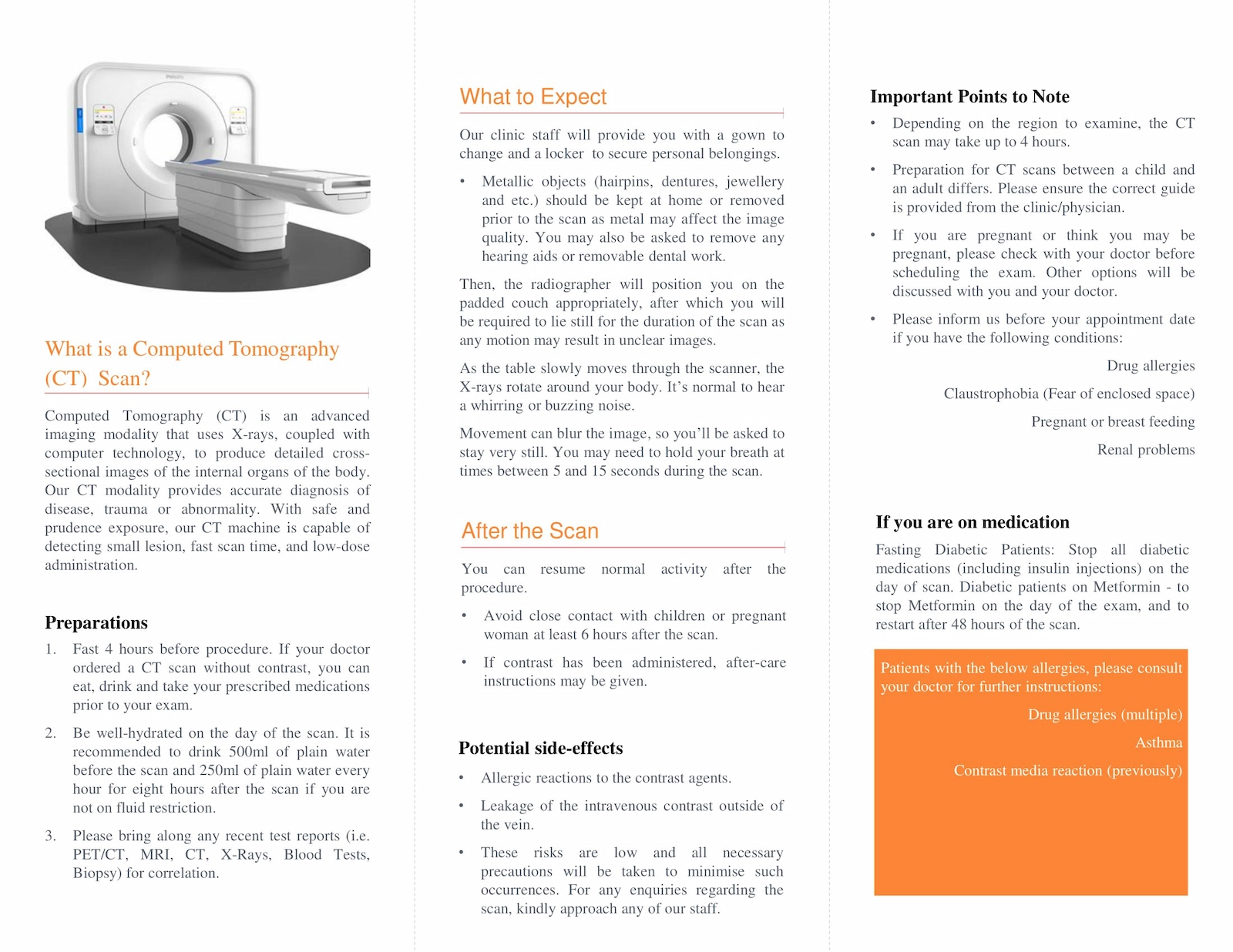
CT scan is an advanced imaging modality that uses x-rays, coupled with computer technology, to produce detailed cross-sectional images of the internal organs of the body. Our CT modality provides accurate diagnosis of disease, trauma or abnormality. Similarly, doctors use MRA images to examine, diagnose and treat blood vessel-related diseases throughout the body. With safe and prudence exposure, our CT machine can detect small lesion, fast scan time, and low-dose administration.
VEO™ - CT750 HD with the latest low dose technology
- VEO™ incorporates an advance image reconstruction process that enables optimal images to be produced at a low radiation dose. It reduces "noise" in the image that obscure detail which may be important for a diagnosis to be made. Radiologists (Doctors who specialize in Diagnostic Imaging) benefit from the clearer, better-defined images, enabling faster and more accurate diagnosis.
- Radiation exposure from CT scan has been a concern for healthcare providers and patients. With VEO™, the patient may be scanned at a dose that is 1/8th that of its predecessor (LightSpeed VCT)
- For example: A regular abdominal CT generates about 6-10 abdominal X-rays worth of radiation. With VEO™ dose reduction technology, the same scan can be achieved at a dose equivalent to only 1 abdominal X-ray.
Features of VEO™:
- Optimal image quality with as little as 1/8 the radiation dose*
- Industry leading spatial resolution (0.23mm) improves analysis of small structures
- Industry leading spatial resolution (0.23mm) improves analysis of small structures
Who will benefit from the low dose technology?
Radiation dose exposure is cumulative and each successive scan increases its detrimental effects. The VEO™ low dose technology is beneficial to anyone who needs to have a CT scan.
It is particularly beneficial to young patients as well as patients requiring repeat CT scans to have as low a radiation dose exposure as possible.
“With VEO™, clearer images are produced at reduced radiation”

Low Radiation Dose
CT abdomen performed at a dose of less than 2 abdominal Xrays*

Low Radiation Dose
CT Heart scan at up to 83% dose reduction

Low Radiation Dose
CT Heart scan at up to 83% dose reduction
What is CT Coronary Angiogram?
- Coronary Artery disease or blockage of arteries in the heart is one of the leading causes of early and sudden death in Singapore.
- CT coronary angiogram can now be performed to evaluate for coronary artery diseases with the advent of new generation CT scanners.
- The CT system is faster and gives higher resolution images compared to previous generation scanners.
- It gives more detailed information to doctors, providing a 3-D computer model of the heart and its arteries.
Magnetic Resonance Imaging (MRI) and MR Angiogram (MRA)
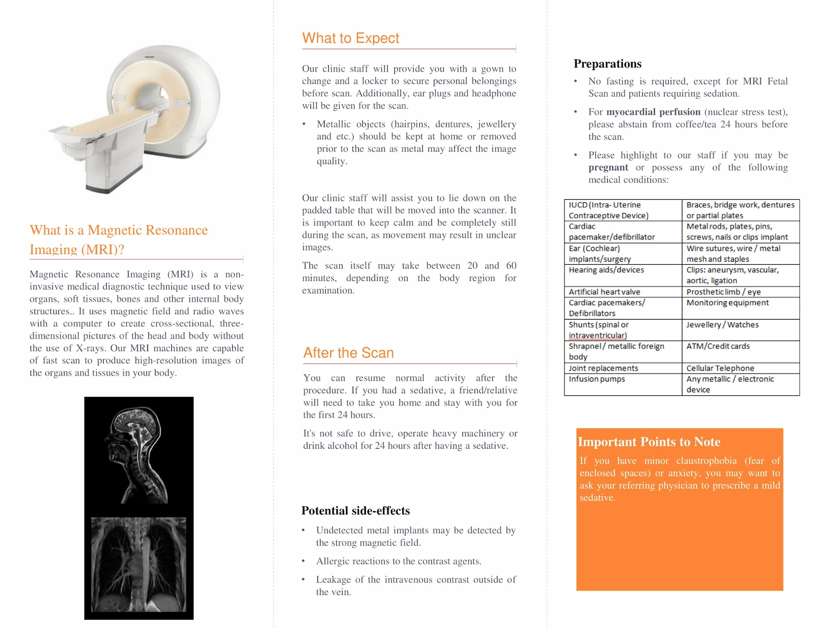
MRI is a non-invasive medical diagnostic technique used to view organs, soft tissues, bones and other internal body structures. It uses magnetic field and radio waves with a computer to create cross-sectional, three-dimensional pictures of the head and body without the use of X-rays. Our MRI machines are capable of fast scan to produce high-resolution images of the organs and tissues in your body. Likewise, for MRA, doctors use the images to examine, diagnose and treat blood vessel-related diseases throughout the body.
What is MRI of the breasts?
- MRI of the breasts is not a replacement for mammography or ultrasound imaging but rather a supplemental tool for detecting and staging breast cancer or other breast abnormalities.
- Recent research has demonstrated that MRI can detect breast lesions that are sometimes missed by mammography and ultrasound.
- MRI can also help detect breast cancer in women with breast implants and younger women with dense breast tissue—both of which are difficult to image using traditional mammography.
- Because MR imaging does not involve X-rays, the procedure is useful to screen women at high risk for breast cancer.
What will I experience during the procedure:
- You will be positioned face down on a moveable bed with your breasts placed into cushioned coils. The bed will then be moved into the magnet of the MRI unit.
- MRI of the breast is a relatively pain-free procedure. Your breasts may feel slightly warm but this is normal and harmless.
- When the contrast agent is injected, it is normal to experience a cooling effect throughout your body.
- You may request earplugs to reduce the noise of the MRI scanner.
- The procedure lasts about 30 minutes.
- Nursing mothers should avoid breastfeeding for 36 to 48 hrs after MRI if contrast material is administered.
Preparation
03
Scan Preparation for Patients
- Mammogram
- MRI
- CT
- US

hould you have other enquiries, please email to imaging@ahppl.com.sg
Thank you!
Connect With Us
Comprehensive care in wellness, aesthetics, imaging, and digestive health — combining
medical expertise with a personal touch. Connect with us to take charge of your health today.
 Asia Health Partners